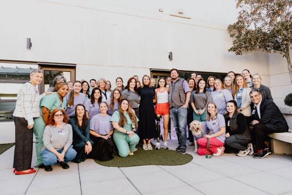
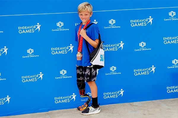
At Ascension St. Vincent’s Southside in Jacksonville, Florida, a local resident regained independence after spine surgery and rehabilitation with care from neurosurgeon Dr. Ali Chahlavi.
Imaging tests, cholesterol tests, heart enzyme blood tests, EKG and blood pressure monitoring help your doctor diagnose heart disease — irregular heartbeat, leaky heart valve, clogged coronary artery and more.
Whether you're experiencing symptoms, managing a heart condition or undergoing a preventive heart screening, our cardiologists work with your primary care doctor to support early detection and personalized treatment planning.
Our cardiology team uses advanced diagnostic tools to identify heart conditions and understand symptoms, helping to create a treatment plan that's right for you.
Your care team provides blood pressure monitoring to help you better understand your heart health and reduce the risk of severe conditions like heart attack, stroke and kidney disease. Whether you're being screened for high blood pressure (hypertension) or managing an existing condition, regular monitoring can keep you healthy. Your heart specialist may recommend doctor visit checks, home blood pressure monitoring or 24-hour blood pressure monitoring.
A blood test is a lab analysis of things that may be found in your blood. You may have blood tests to keep track of how well you are managing a condition, such as diabetes or high cholesterol. You may also have them for routine checkups or when you are ill.
This is also known as a cardiac or heart cath. For this test, your doctor guides a small catheter (hollow tube) through the large artery in your upper leg, or sometimes your wrist or arm, into your heart. This procedure lets your doctor take a close look at the heart to identify concerns and to perform other tests or procedures.
This imaging procedure uses an X-ray machine and a computer to create 3-D pictures of the heart. Sometimes a dye is injected into a vein so that your heart arteries can also be seen. Sometimes medicine is given to lower your heart rate so it captures a better image. It can also be used to find out how much calcium is in your heart arteries. Calcium is a marker for coronary artery disease.
This procedure uses a combination of large magnets, radio waves, and a computer to make detailed images of organs and structures in your body. Your doctor may order an MRI of the heart to look at the heart valves and major vessels. It can also detect coronary artery disease and how much damage it has caused. It can also assess heart problems that have been present since birth. It can find tumors and other conditions. Your doctor may order this test before other procedures such as angioplasty or stenting of the coronary arteries and heart or vascular surgery.
This test is done during a cardiac catheterization. For this test, your doctor guides a small catheter (hollow tube) through the large artery in your upper leg, or sometimes your wrist or arm, into your heart. Dye is given through the catheter, and moving X-ray pictures are taken as the dye travels through your heart arteries and heart chambers. This comprehensive test shows narrowing in the arteries, heart chamber size, how well your heart pumps, and how well the valves open and close. It also measures the pressures within the heart chambers, arteries, and veins.
This test is a CT scan that measures the amount of calcium in the walls of your coronary arteries. Buildup of calcium, or calcifications, are signs of atherosclerosis or coronary heart disease.
Your doctor may recommend this test if you have a condition that involves the blood vessels. CT angiography is a type of medical test that combines a CT scan with an injection of a special dye. This is to make pictures of blood vessels and tissues in a part of your body. The dye is injected through an IV (intravenous) line started in your arm or hand. A CT scan is a type of X-ray that uses a computer to make images of your body. The dye injected to do CT angiography is called a contrast material. This is because it highlights the blood vessels and tissues being studied.
It's used to check the heart's function and structures. During the procedure, a transducer (like a microphone) sends out sound waves at a frequency too high to be heard. When the transducer is placed on the chest at certain locations and angles, the sound waves move through the skin and other body tissues to the heart tissues. The waves bounce or "echo" off the heart structures. These sound waves are sent to a computer that can create moving images on the screen of the heart walls and valves.
This test records the electrical activity of the heart, shows abnormal rhythms (arrhythmias), and can sometimes detect heart muscle damage.
For this test, insulated electric catheters are placed through the large vein in the upper leg and threaded into the heart. It's used to test the heart's electrical system. It helps your doctor look at what might be causing abnormal heart rhythms.
For this test, you wear a small, portable, battery-powered machine used to record ECG over several weeks. Each time you have symptoms, you press a button on the recorder to record the ECG sample. As soon as possible, you will transmit this sample to your doctor for evaluation.
For this test, you wear a small, portable, battery-powered ECG machine. Small patches (wired electrodes) are attached to the skin over your heart. The monitor records heartbeats over a period of 24 to 48 hours during normal activities. At the end of the time period, you will return the monitor to your doctor so it can be read and evaluated. Some Holter monitors can be worn for up to 2 weeks. These monitors are patches and don't require wires.
This is a nuclear scan that helps your doctor understand the flow of blood through the coronary arteries to the heart muscle.
This is also called a treadmill or exercise ECG. This test is done to monitor the heart while you walk on a treadmill or pedal a stationary bike. Your doctor also monitors your breathing and blood pressure. A stress test may be used to detect coronary artery disease, or to determine safe levels of exercise after a heart attack or heart surgery. This test can also be done using special medicines that stress the heart in a similar manner as exercise does. Sometimes a stress test will collect ECG information along with heart ultrasound pictures. This is called an exercise or stress echocardiogram (echo). It's more sensitive and specific than ECG stress testing alone.
This test is similar to a transthoracic echocardiogram. But it's done with medicine to help you relax (sedation). It's considered invasive because a probe is put into your body. In this test, you will swallow a small probe about the size of your thumb. The probe passes down the esophagus, which lies directly behind the heart. It allows a much closer look at the heart's structure and function than a standard echocardiogram done on the skin of the chest. It can better look at heart valve structure and function. Your doctor can better see any blood clots that may be in the heart.
A heart scan is a quick, painless way to detect early signs of heart disease before symptoms appear. If you have risk factors like high blood pressure, high cholesterol, or a family history, this simple test could prevent a heart attack. Take control of your heart health today.
At Ascension St. Vincent’s Southside in Jacksonville, Florida, a local resident regained independence after spine surgery and rehabilitation with care from neurosurgeon Dr. Ali Chahlavi.
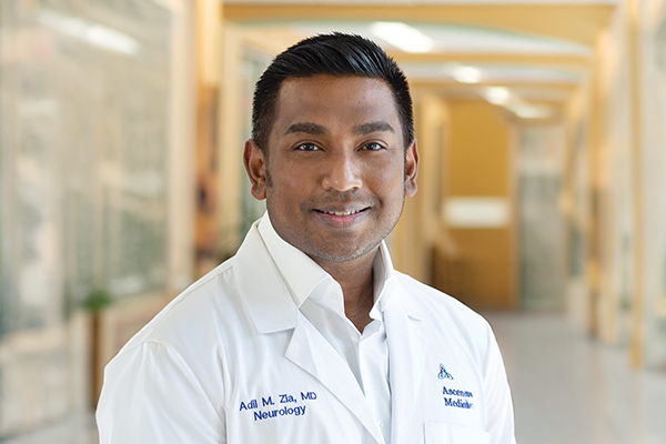
Adil Zia, MD, neurologist and stroke director at Ascension St. Vincent’s hospitals in Jacksonville, Florida, answers questions about stroke signs and symptoms.

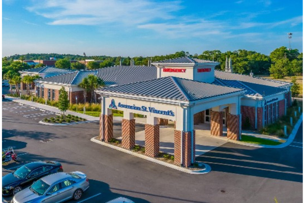
Ascension St. Vincent’s has opened another freestanding emergency department on Jacksonville’s Southside to serve its growing community.
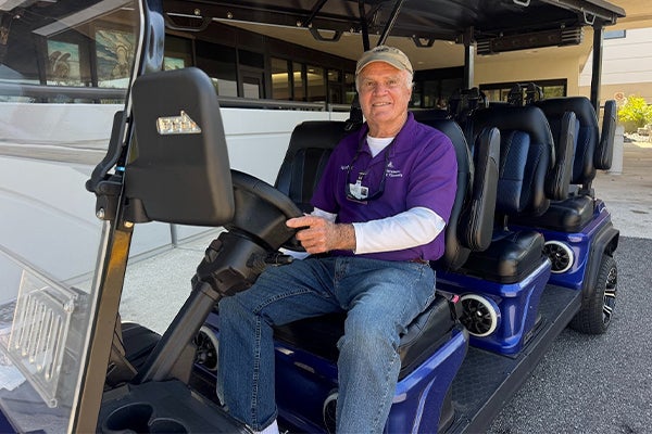
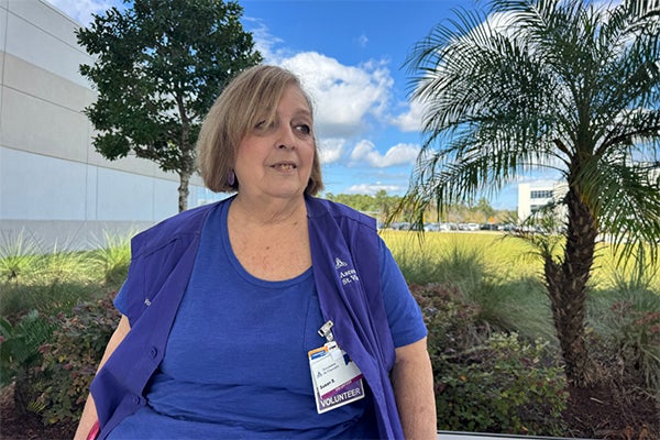
At Ascension St. Vincent’s Southside, a dedicated volunteer golf cart driver transforms parking lot arrivals into compassionate, accessible care, even securing new transportation through a grant.
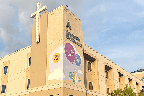
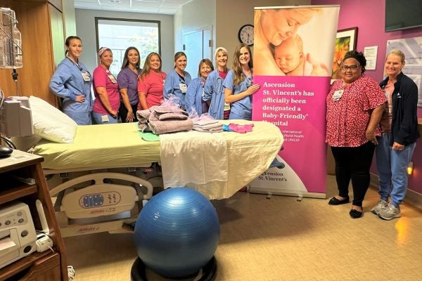
Ascension St. Vincent’s Clay County Level II NICU expansion is a step toward bringing specialized newborn care closer to home for families in Northwest Florida.
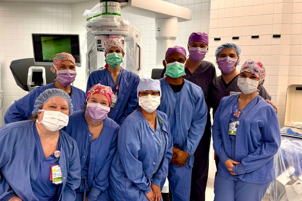
Ascension St. Vincent’s Riverside in Jacksonville, Florida, added a new surgical system that expands surgical care options for patients across several specialties.
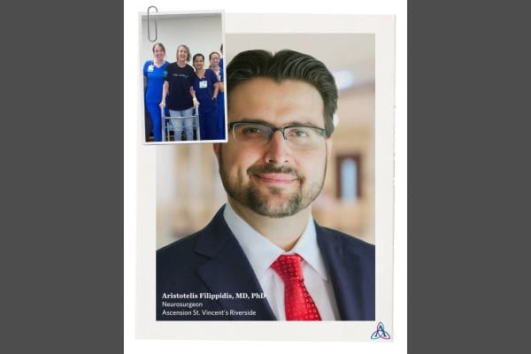
During a July family wedding trip, a mother of the bride received lifesaving neurosurgical care for a brain tumor from Dr. Aristotelis Filippidis at Ascension St. Vincent’s Riverside.
February 27, 2026
After a routine gynecology visit revealed stage three cervical cancer, Kristina began treatment with Martin Martino, MD, gynecologic oncologist at Ascension St. Vincent’s Riverside in Jacksonville, Florida.

February 24, 2026
Ascension Rx Southside Pharmacy is an accredited specialty pharmacy in Jacksonville, Florida that provides advanced medication support and personalized care to local residents.
February 23, 2026
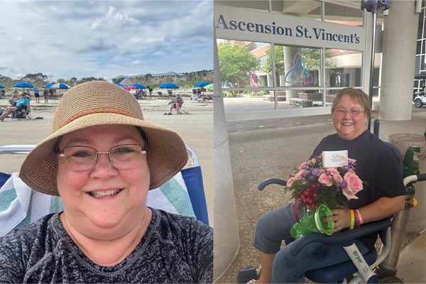
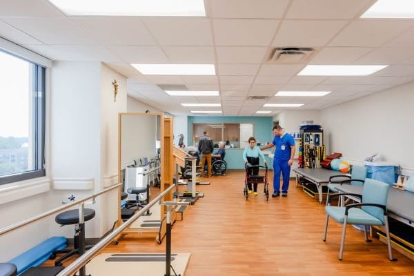
At Ascension St. Vincent’s Southside Inpatient Rehabilitation specialists help you get back to doing what you love.
February 19, 2026
When a Jacksonville man chose a freestanding ER like Ascension St. Vincent’s Emergency Care - Arlington, for emergency care he had access to life-saving care close to home.
February 5, 2026
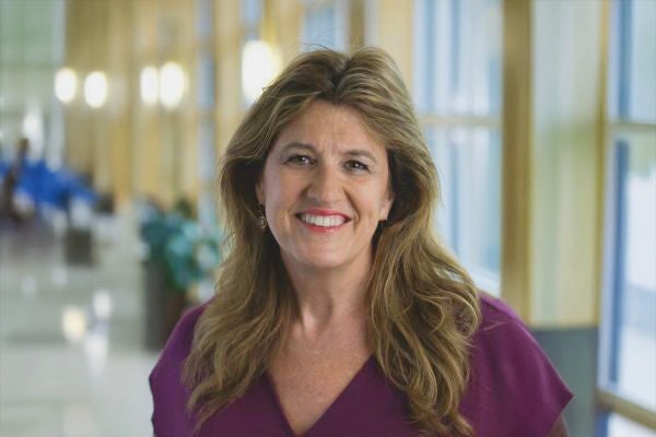
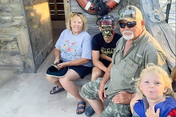
In September 2025, Sherri received lifesaving cardiac care from Dr. Lucien Abboud at Ascension St. Vincent’s Cardiology – St. Johns after testing revealed a serious heart blockage.

February 4, 2026
When illness or injury happens, understanding when to visit an Ascension St. Vincent’s ER or urgent care can potentially save your life.

January 29, 2026
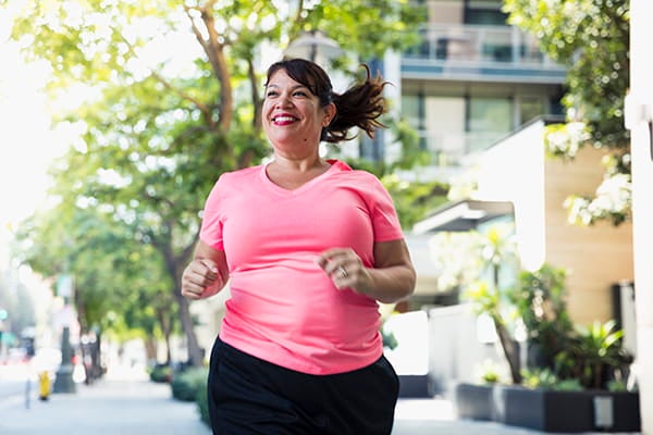
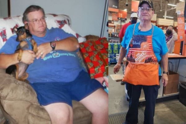
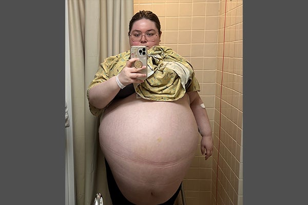
Ascension St. Vincent’s bariatric program in Jacksonville, Florida, shares some truths about weight-loss options.
January 12, 2026
An Ascension St. Vincent’s St. Johns County hospital volunteer transformed her grief from losing her husband into a mission of kindness and compassion, inspired by the care her late husband received.
December 8, 2025
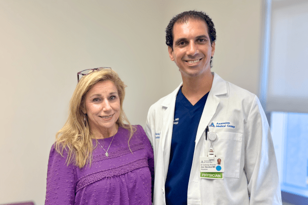
After surviving a lightning strike in 2025, Georgia teacher Angela received AFib care from Dr. Amr Barakat at Ascension St. Vincent’s Riverside in Jacksonville, Fl.
December 1, 2025
Bariatric Medical Director Miroslav Uchal, MD at Ascension St. Vincent’s shares the new non-surgical weight-loss options for Jacksonville, Florida residents.
November 21, 2025
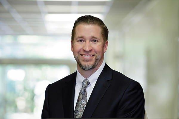
Dr. Saumil Oza, cardiologist at Ascension St. Vincent’s Riverside in Jacksonville uses a combined procedure to treat patients with AFib to help with faster recovery and reduce their risk for stroke.
October 28, 2025
At Ascension St. Vincent’s in Jacksonville, Dr. Martin Martino helped an 88-year-old woman from Orange Park overcome uterine cancer through compassionate, personalized care and advanced treatment.
October 28, 2025
At Ascension St. Vincent’s Riverside in Jacksonville, Florida, neurosurgeon Dr. Aristotelis Filippidis performed brain tumor surgery that saved a patient’s life.
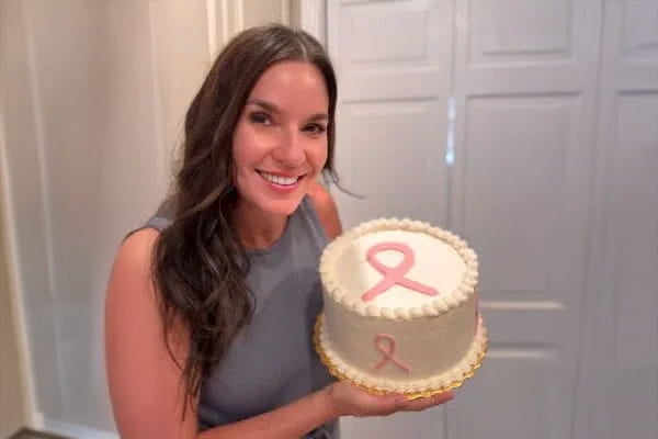
Patient thanks doctor at Ascension Sacred Heart Surgical Breast Oncology Bay in Panama City for her guidance and care during her breast cancer journey.
October 20, 2025
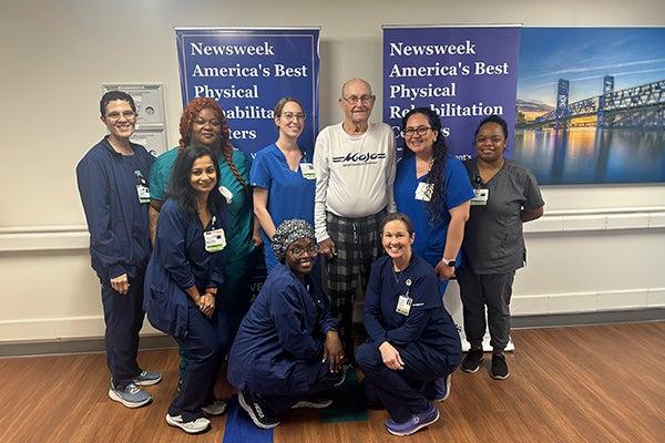
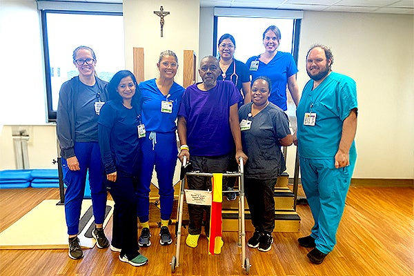
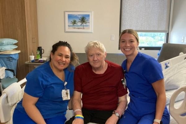
After a spine cancer diagnosis, Mr. Santorelli turned to Dr. Stephen Parker, neurosurgeon, and the care team at Ascension St. Vincent’s in Jacksonville, Florida, for spinal surgery and rehabilitation to find relief from pain and regain strength and independence.
Deborah A. Weyer, MD, pediatrician at Ascension St. Vincent’s in Jacksonville, Florida, is helping children and families prevent and manage obesity through early screenings, healthy lifestyle guidance, and personalized pediatric care.
Neurosurgeon at Ascension St. Vincent’s Riverside in Jacksonville, FL, is using new technologies to care for patients with brain and spine tumors.
July 9, 2025
Ascension St. Vincent’s Southside Inpatient Rehabilitation Facility in Jacksonville, Florida, helps stroke patients recover and regain their strength after a hospitalization.

If incidents occur this Fourth of July, knowing where and when to get immediate and emergency care is important.
June 23, 2025
Ascension urgent care and orthopedic teams are here to help you manage your summer injury.
Ascension St. Vincent’s Southside Inpatient Rehabilitation Facility in Jacksonville, Florida, helps stroke patients recover and regain their strength after a hospitalization.

Mobile health nurse Peggy Kennon shares the importance of giving care to communities with limited access to health care through her work at Ascension St. Vincent’s in Jacksonville, Florida.
April 30, 2025
Ascension Rx medication assistance team at Ascension Sacred Heart in Pensacola, Florida, collaborated with a missionary worker diagnosed with brain cancer to overcome the costs of medication allowing her to focus on her recovery.
April 22, 2025
Finding Strength and Support at Ascension St. Vincent’s Southside Inpatient Rehabilitation Facility

Dr. Augustus Perez, a pediatric neurosurgeon at Ascension Sacred Heart in Pensacola, Florida, helps an infant boy with a developmental condition called craniosynostosis.
Alabama couple finds compassionate care through every step of their journey at Ascension Sacred Heart in Pensacola, during a high-risk triplet pregnancy and delivery.
February 25, 2025
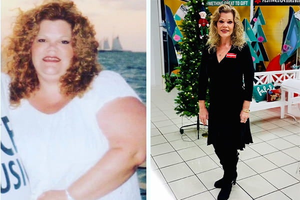
After having bariatric surgery, Jacksonville grandmother credits Ascension St. Vincent’s weight loss program for helping her with her weight-loss journey.
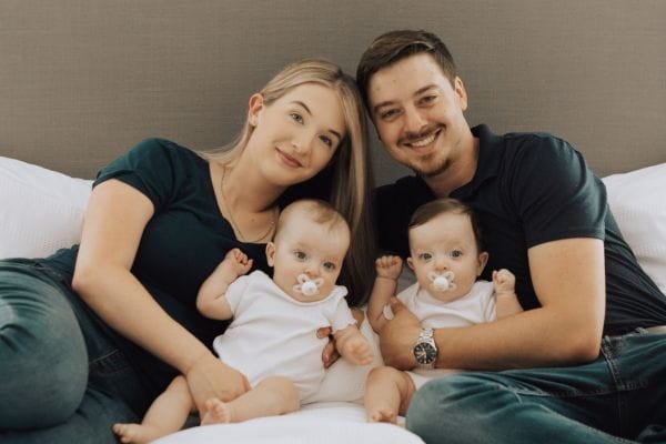
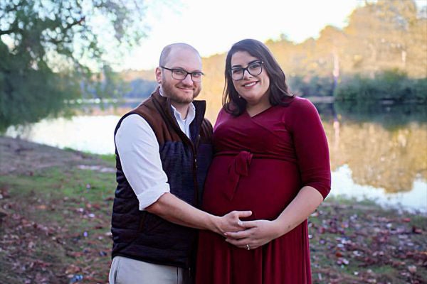
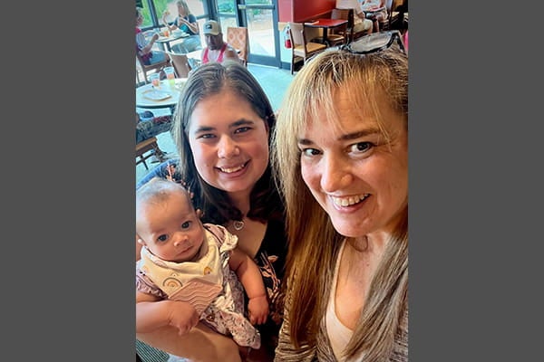
An executive assistant at Ascension St. Vincent’s Southside received advanced maternity care for her high-risk twin pregnancy in Jacksonville, Florida.
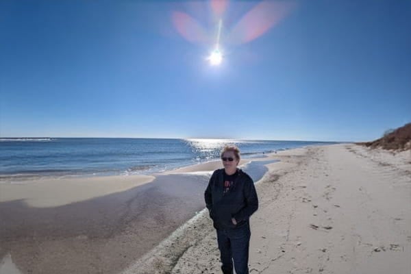
Teen reunites with 70 care team members who played a critical role in her journey to recovery after surviving a shark attack at Rosemary Beach, FL.
November 13, 2024
Ascension St. Vincent’s St. Johns County nurse manager’s honors the hospital's 100-years- of service by starting a food pantry for the St. Johns, FL, community.
Ascension St. Vincent’s Clay County rehabilitation team worked together to support the patient's journey to recovery during more than 100 days in the hospital in Middleburg, FL.

November 5, 2024
Ascension St. Vincent's Riverside OR nurse’s personal experience with having family members living with heart disease inspired her to raise money for heart research in Jacksonville, Florida.
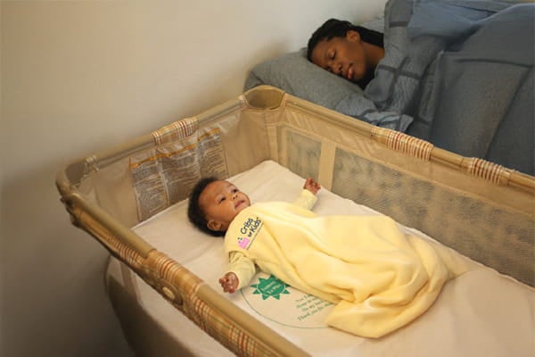
In Jacksonville, Florida, expecting families learn ways to prevent infant sleep-related deaths at Ascension St. Vincent’s
Deborah A. Weyer, MD, pediatrician at Ascension St. Vincent's Pediatrics in Lake Asbury, Florida shares tips for parents to keep their children physically and mentally healthy during the school year.
Orthopedic surgeon, Dr. Mark Elliott helps a former hospice nurse find pain relief with a shoulder repair surgery at Ascension Sacred Heart in Pensacola, FL.
Justin Labrato shares what he has learned while serving as the Chief Operating Officer at Ascension Sacred Heart Medical Group in Pensacola, FL.
February 26, 2024
Ascension St. Vincent’s Riverside gynecologic oncology surgeon in Jacksonville, FL, helps patient balance surgical care with religious beliefs

January 31, 2024
Ascension Sacred Heart surgeons pay it forward by mentoring future health professionals in Pensacola, FL.
January 10, 2024
Ascension St. Vincent’s Clay County’s maternity care in Middleburg, Florida ranked among the nation’s best
October 12, 2023
A well-known patient advocate in Pensacola, FL, overcame breast cancer twice with early detection and treatment at Ascension Sacred Heart.
Nurse practitioner Heather Ledford at Ascension St. Vincent’s travels to patients’ homes to give personalized care when they are unable to leave the house.
September 13, 2023
Pensacola pediatric trauma director at Studer Family Children’s Hospital applauds new golf cart law that raises driving age.
September 1, 2023
Pensacola man doesn’t delay seeking care at Ascension Sacred Heart when his skin starts to discolor from pancreatic cancer.
August 31, 2023
Wear and tear of the hip, knee and other joints is part of aging, but Pensacola, FL patients find relief with minimally invasive surgery at Ascension Sacred Heart.
August 25, 2023
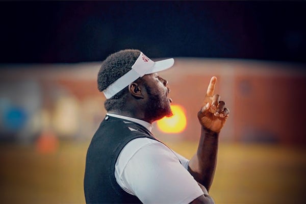
Pensacola football coach suffers stroke mid-game, receives lifesaving treatment at Ascension Sacred...
August 24, 2023
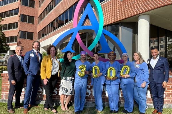
The 300th TAVR patient at Ascension Sacred Heart in Pensacola, FL, is happy with his improved quality of life after surgery.
August 23, 2023
Ascension Sacred Heart ER nurse helps son in an incident that left him partially amputated in Pensacola, FL.
August 21, 2023
Ascension St. Vincent’s faith community nurse in Jacksonville, FL shares how she was called to serve.
August 18, 2023
Dr. Kurt T. Stockamp, general surgeon at Ascension Sacred Heart in Pensacola, Florida answers questions related to gallbladder surgery.
August 16, 2023
Ascension Sacred Heart has earned David’s trust over the years. From primary care to orthopedics to cardiology, he says he has confidence in his care team. When he felt chest pain, fatigue and dizziness, he says he knew where to turn.
July 31, 2023
Jennifer Morton, DNP, RN, takes on a new leadership role at Ascension as the Senior Director of Nursing Practice and Operations for the organization.
Florida vacationers receive emergency care from Ascension Sacred Heart Pensacola in Pensacola, FL, after an unexpected cardiac event.
Ascension St. Vincent’s new technology gives Jacksonville man a second chance with real-time management of heart failure.
Dr. Amr Barakat, cardiac electrophysiologist at Ascension St. Vincent's Cardiology in St. Johns County, Florida answers questions about treating AFib.
Patient returns to serving others after having the WATCHMAN procedure at Ascension Sacred Heart Bay in Panama City, FL.
A Pensacola, Florida man embraces a new active life after having weight-loss surgery and support at Ascension Sacred Heart.
June 1, 2023
Dr. Lauren Stipp, cardiologist at Ascension Medical Group Sacred Heart Cardiology 30A in Watersound, FL answers common questions about heart CT scans.
Dr. Jairan L. Duke-Elmore, obstetrician at Ascension Medical Group Sacred Heart in Pensacola, Florida answers common questions about pregnancy and heart disease.
May 31, 2023
Ascension St. Vincent’s Inpatient Rehabilitation Unit in Jacksonville, FL helped this grandmother regain strength and function in her muscles.
May 17, 2023
Dr. Marcia Schmidt, gynecologic oncologist at Ascension Medical Group St. Vincent’s Gynecologic Oncology and Advanced Women’s Health in Jacksonville, Florida answers common questions about cervical cancer.
Dr. Shailee Shah, cardiologist at Ascension Medical Group St. Vincent’s Women’s Heart and Prevention Clinic in Saint Johns, Florida answers common questions about heart disease in women.
Weighing just a pound-and-a-half and stretching the length of a ruler, micro-preemie Teigen shocked her mother by making an early entrance at 23 weeks old at Studer Family Children’s Hospital at Ascension Sacred Heart last year.
May 9, 2023
How one question changed nurse's career path
As a Neonatal Intensive Care Unit nurse caring for the tiniest patients at Studer Family Children’s Hospital, Chelsea Fredrickson’s life has come full circle.
After having minimally invasive hip replacement, patient feels 20 years younger.

April 20, 2023
Dr. Maria Lahti, cardiologist at Ascension Medical Group Sacred Heart Cardiology Destin in Destin, Florida answers common questions about heart disease.

April 20, 2023
Dr. Sara Largen, pediatrician at Ascension St. Vincent’s in Jacksonville, FL, shares what to expect at your child’s appointments.
April 20, 2023
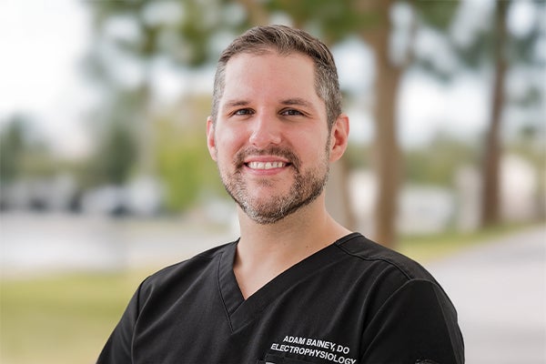
Dr. Adam Bainey, cardiac electrophysiologist at Ascension Sacred Heart in Miramar Beach, Florida answers your common questions about AFib treatment.
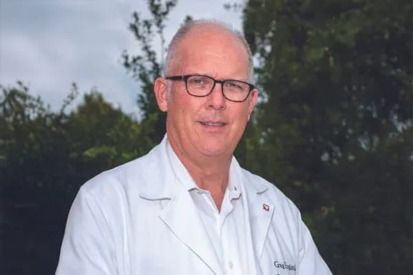
Dr. Greg England, cardiothoracic surgeon at Ascension Sacred Heart Bay in Panama City answers common questions about heart surgery.
February 7, 2023
One month after Allison Fisher had a 104-lb. ovarian tumor removed at Ascension St. Vincent’s Riverside, she feels she has a second chance at life.

After suffering an incomplete spinal cord injury seven years ago, recurring urinary tract infections have taken a toll on his health.
January 17, 2023
One morning seven years ago, Angela Lane considered canceling her annual mammogram at the Ann L. Baroco Center for Breast Health and Mammography at Ascension Sacred Heart.